How Do You Know if a Cat With Cutaneous Mast Cell Tumors Has Visceral?
What is a mast cell?
A mast prison cell is a type of white blood cell that is institute in many tissues of the body. Mast cells are allergy cells and play a role in the allergic response. When exposed to allergens (substances that stimulate allergies), mast cells release chemicals and compounds, a process called degranulation. One of these compounds is histamine. Histamine is virtually commonly known for causing itchiness, sneezing, and runny eyes and nose – the common symptoms of allergies. But when histamine (and the other compounds) are released in excessive amounts (with mass degranulation), they tin can cause full-torso effects, including anaphylaxis, a serious, life-threatening allergic reaction.
What is a mast cell tumor?
A mast cell tumor (MCT) is a blazon of tumor consisting of mast cells. Mast cell tumors can form nodules or masses in the skin (and other organs), and cause enlargement of the spleen and intestine. Mast jail cell tumors are the virtually common splenic tumor (tumor of the spleen), 2nd nigh common skin tumor, and third about common abdominal tumor in cats.
What causes this cancer?
Why a particular true cat may develop this, or any cancer, is by and large not straightforward. Very few cancers take a single known crusade. Most seem to be caused by a complex mix of risk factors, some environmental and some genetic or hereditary. A genetic mutation in a protein involved in the replication and division of cells (called KIT) has been well-described in the development of MCTs in dogs. In cats, virtually 67% of MCTs likewise have this mutation.
What are the signs that my cat may have a mast cell tumor?
Most mast prison cell tumors are seen as house plaques (difficult, flattened areas) or nodules (small lumps) in the skin. The head and neck regions are the near usually affected areas, peculiarly the top of the head and either or both ears. There may be itching because the tumors produce substances that cause inflammation.
If your true cat has the splenic form of the disease, the most commonly observed signs are weight loss, vomiting, and loss of ambition. This is a outcome of the compounds released by the cancer that make your cat feel sick.
"If your cat has the splenic course of the disease, the most commonly observed signs are weight loss, airsickness, and loss of appetite."
The intestinal grade, depending on how severe the affliction is, may cause vomiting, diarrhea, fresh red blood in the stool, or black/tar-colored stool (the discoloring evidence of digested claret). In some cases, your veterinarian may be able to feel a mass within the abdomen during a concrete examination.
How is this cancer diagnosed? 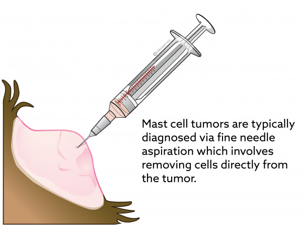
Typically, this cancer is relatively easily diagnosed by cytology. Cytology is the examination of cells under a microscope. These cells are retrieved past placing a needle into the mass, suctioning out some cells, and placing them on a microscope slide. This procedure is chosen a fine needle aspiration (FNA). A veterinary pathologist then examines the slide nether a microscope.
In cases that are questionable, surgical excision of a piece of the tumor (a biopsy) or the unabridged tumor tin be performed. Pieces of the tumor will so be examined under the microscope. This is called histopathology. Histopathology gives the pathologist much more information about the type of tumor, how ambitious information technology is, and the margins (the border between healthy and cancerous tissue). If your veterinarian submits the unabridged tumor to the pathologist, the likelihood that the cancer has been fully removed tin can be determined.
How does this cancer typically progress?
There are iii distinct forms or syndromes of MCT in the cat:
Cutaneous MCT
Siamese cats appear to exist predisposed to this course of MCT. Based on how the cells appear under the microscope – well-differentiated (i.east., what a more normal mast jail cell looks like) or poorly-differentiated (i.e., what a very aberrant mast cell looks like) – the illness progression and prognosis may vary. The well-differentiated tumors tend to act less aggressively. Staging (searching for potential spread to other locations in the body) should also be pursued. This may include bloodwork, urinalysis, 10-rays of the lungs, and maybe an abdominal ultrasound. If any lymph nodes are enlarged or feel aberrant, further sampling may exist pursued to determine if spread is present. Some cats with cutaneous MCT will take aberrant mast cells in their spleen, but can have them elsewhere also. Staging, therefore, is always recommended.
Splenic/visceral (associated with internal organs) MCT
The spleen is a filtering organ that contains cerise blood cells and white blood cells (including mast cells). Approximately 15% of cats with abnormal or diseased spleens are diagnosed with splenic MCT. This cancer has the potential to spread to other organs as well (e.one thousand., the liver, lymph nodes, bone marrow, lungs, and less unremarkably, the intestines).
Abdominal MCT
Intestinal MCT typically involves the small intestine, but in that location are some reports citing MCT of the colon every bit well. Unfortunately, abdominal MCT commonly spreads to neighboring organs and lymph nodes. When this happens, some cats will develop fluid in their abdomen (called an effusion).
What are the treatments for this blazon of tumor?
Surgical removal of the mass(es) is the treatment of choice whenever possible. Depending on the findings with histopathology and staging, chemotherapy may be suggested. In some cases, if the mass is non completely removed (meaning some cancerous cells are left backside) or in a location that makes surgery as well difficult or risky for your cat, radiation therapy may be suggested.
"Depending on the findings with histopathology and staging, chemotherapy may be suggested."
In cases of intestinal MCT and MCT involving the spleen, surgery is also the handling of choice.
All three forms of mast cell tumors can release compounds that increase acid product in the tum, causing breadbasket upset and heartburn-like symptoms. Your veterinarian may prescribe anti-nausea and antacid medications to aid your cat feel better.
Is there anything else I should know?
Preventing your cat from rubbing, scratching, licking, or biting the skin tumor(southward) volition reduce itching, inflammation, ulceration, infection, and bleeding. Whatever ulcerated areas need to be kept make clean.
Later surgery, the surgical site needs to be kept clean and your cat should not exist allowed to lick or chew at the site. Your veterinarian may recommend the employ of an Elizabethan collar (East-collar or cone). Be sure to report whatever loss of sutures or significant swelling or haemorrhage to your veterinarian. If you require additional advice on post-surgical care, please ask your veterinary healthcare squad.
Source: https://vcahospitals.com/know-your-pet/mast-cell-tumors-in-cats
0 Response to "How Do You Know if a Cat With Cutaneous Mast Cell Tumors Has Visceral?"
Post a Comment